The World’s Smallest CBCT, With a Surprisingly Big Perspective.
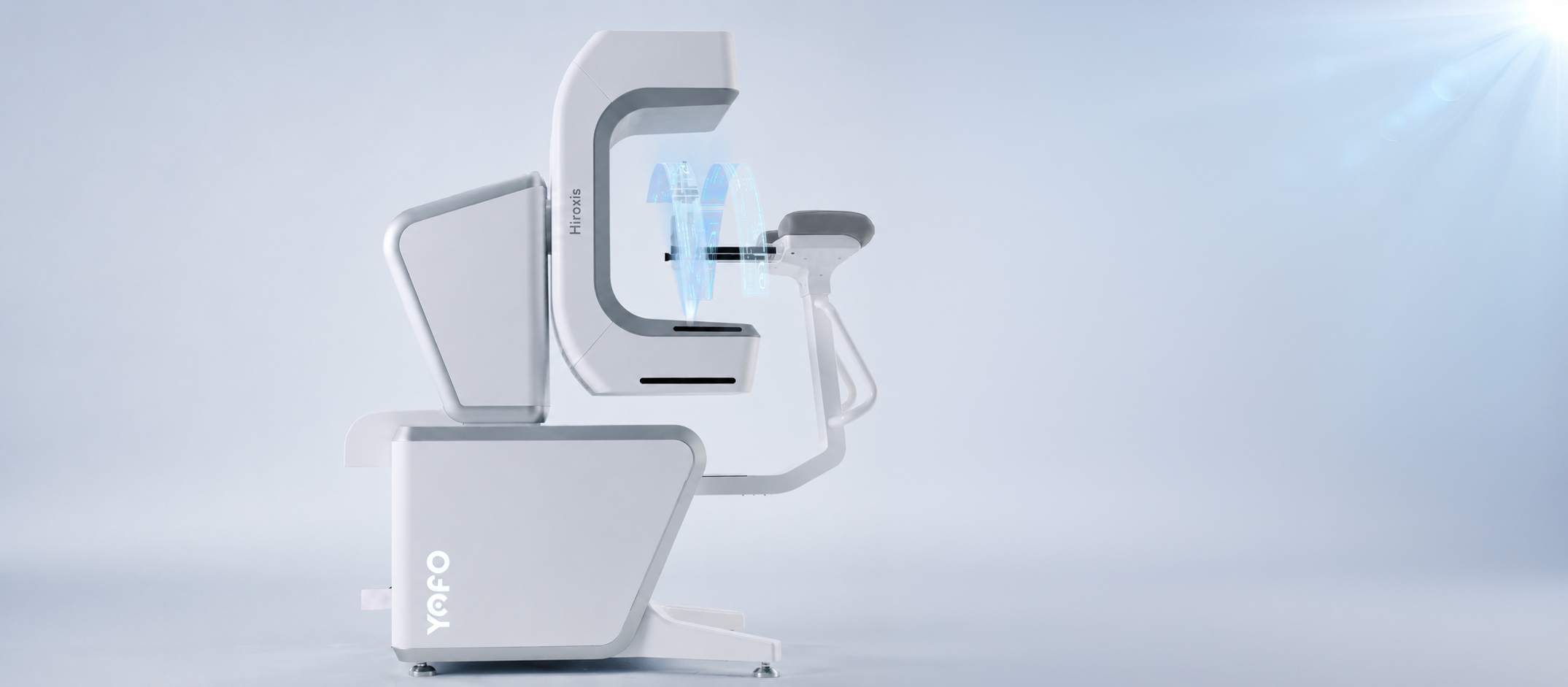
Designed to bring advanced imaging performance to practices of any size, Hiroxis pairs the world’s smallest CBCT footprint with a field of view of up to 16 × 15 cm and simultaneous multiple-view imaging without image stitching, powered by the world’s first helical dental CBCT technology.
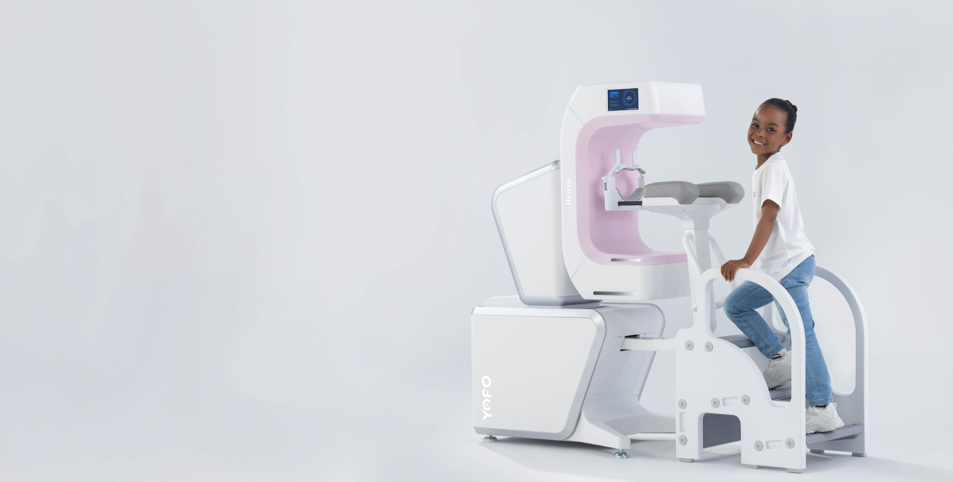
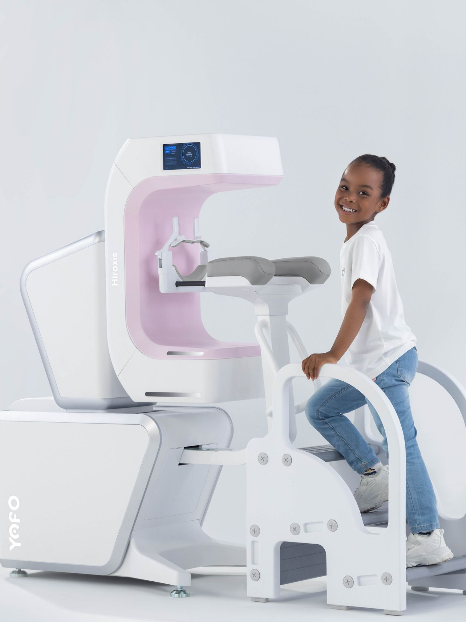
Ready straight out of the box, Hiroxis requires no installation and no calibration. Its shoulder rests enhance patient stability, helping reduce motion and improve
scan consistency.
Ready Out of the Box.
No Installation. No Calibration.
Fits Practices of Any Size.
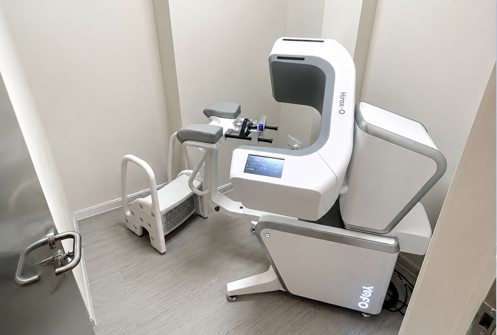
Simply unpack, connect and start scanning.
Advanced Imaging Beyond Its Size
Powered by the world’s first helical dental CBCT technology, Hiroxis delivers broad anatomical coverage from an exceptionally compact system.
Wide Coverage. Sharp Clarity.
One Scan. Everything.
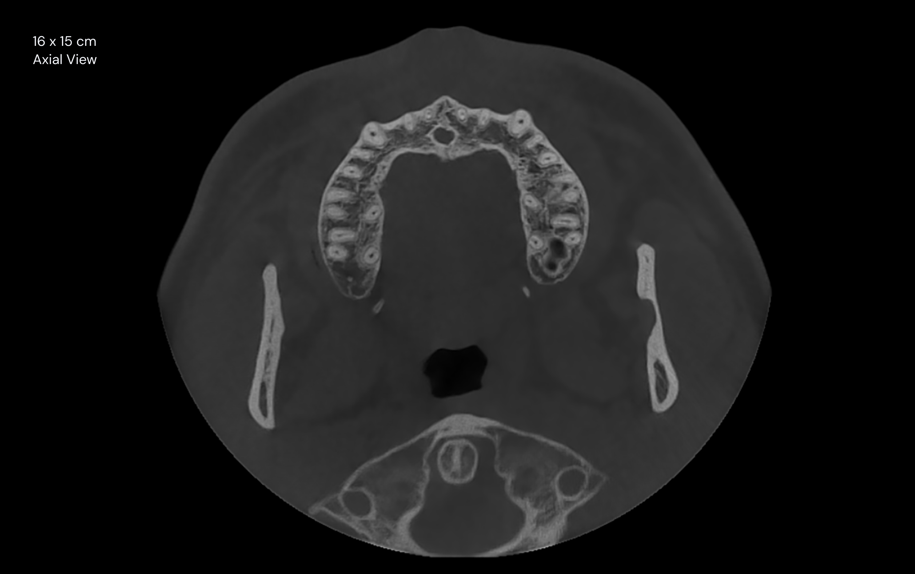
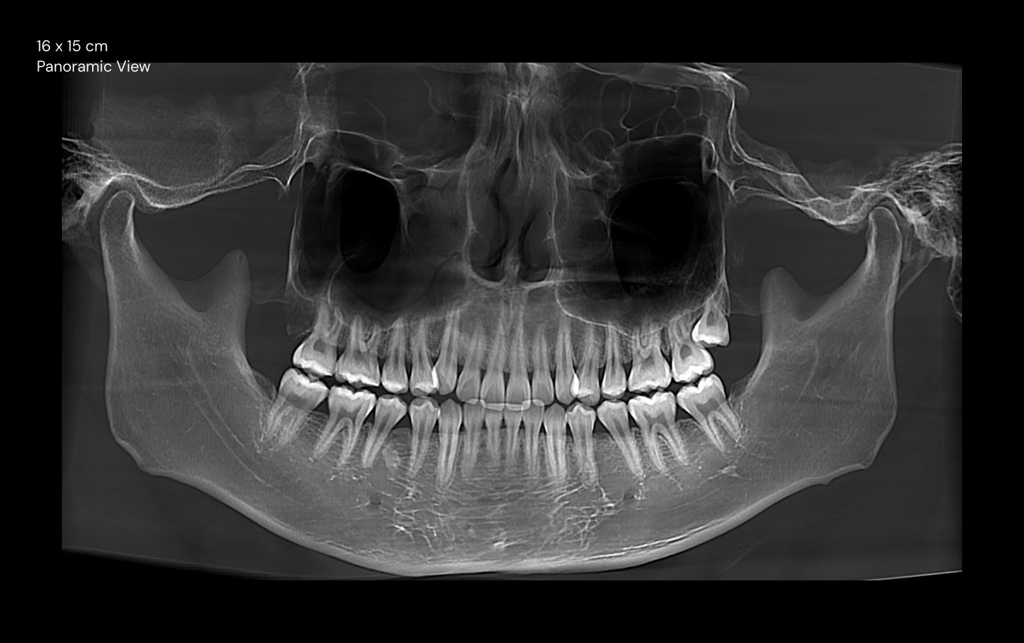
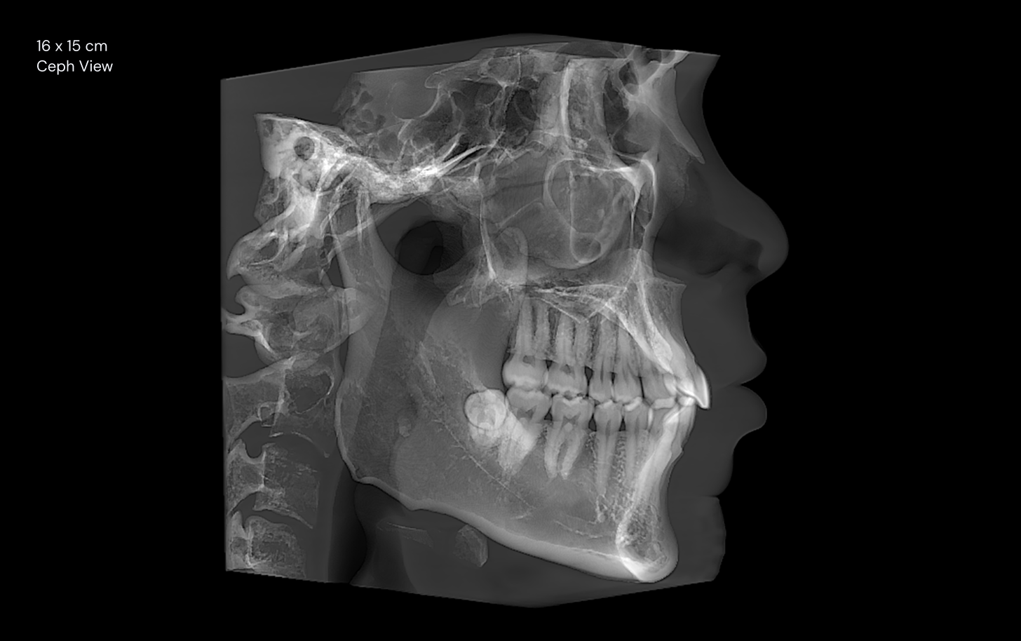
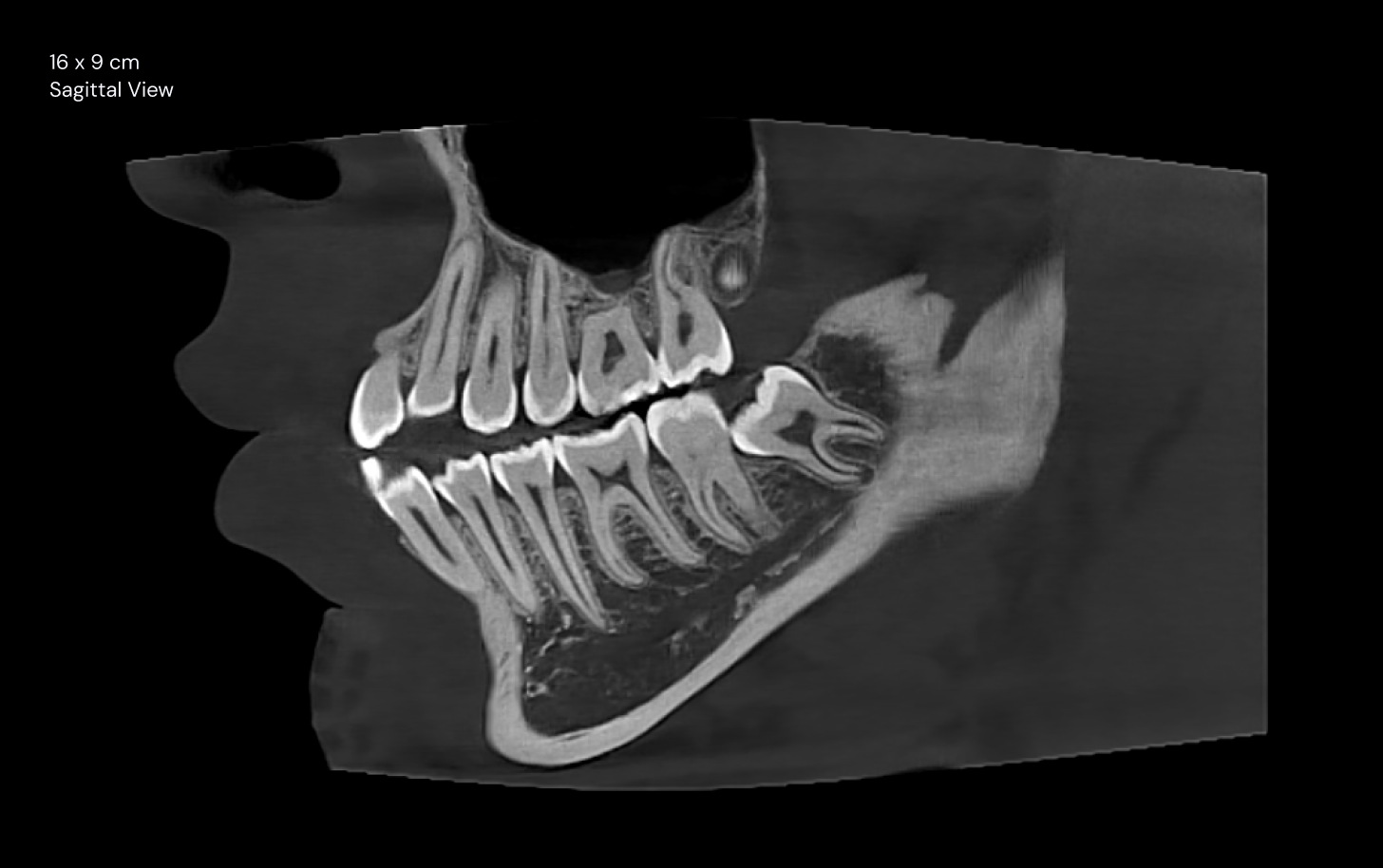
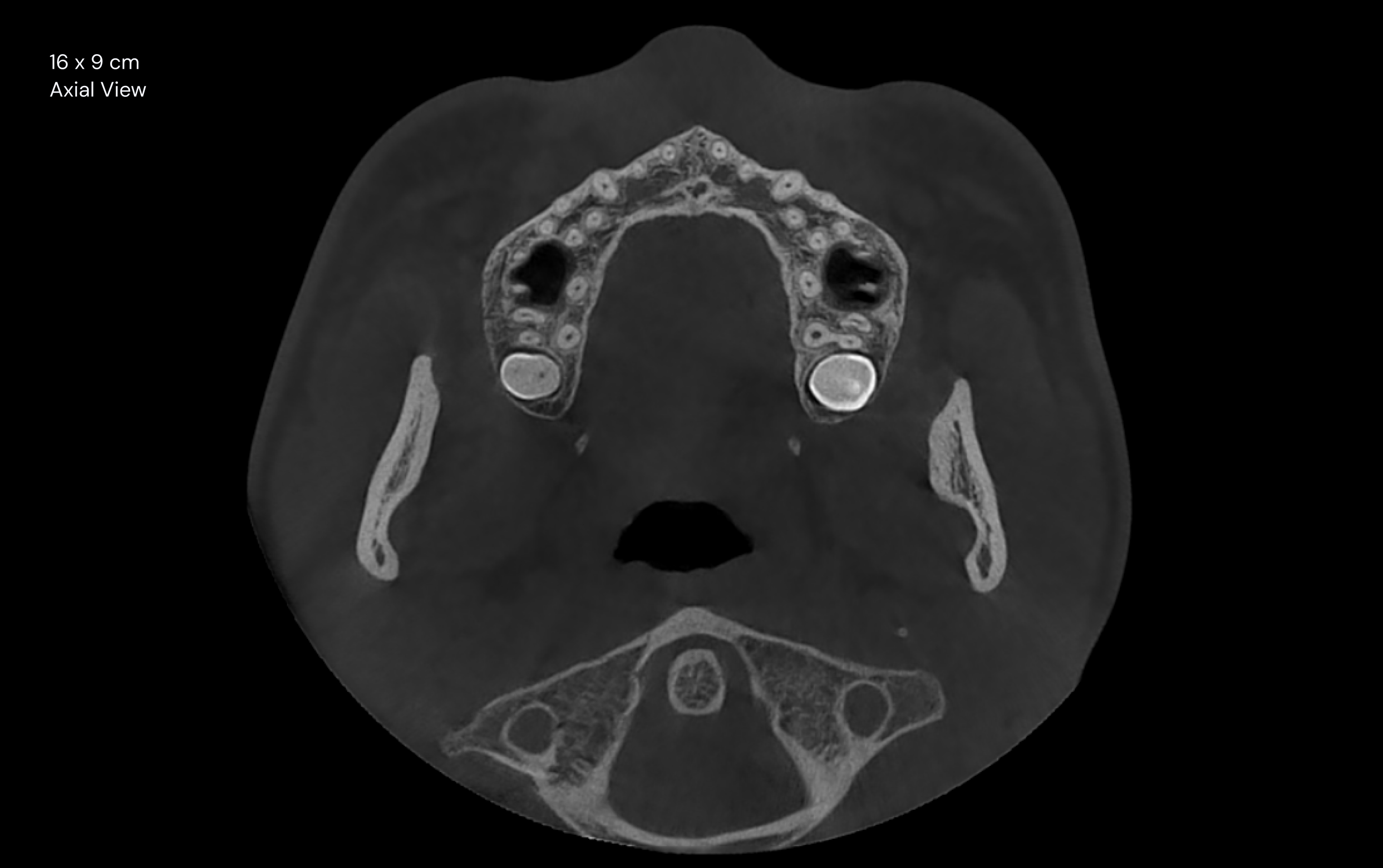
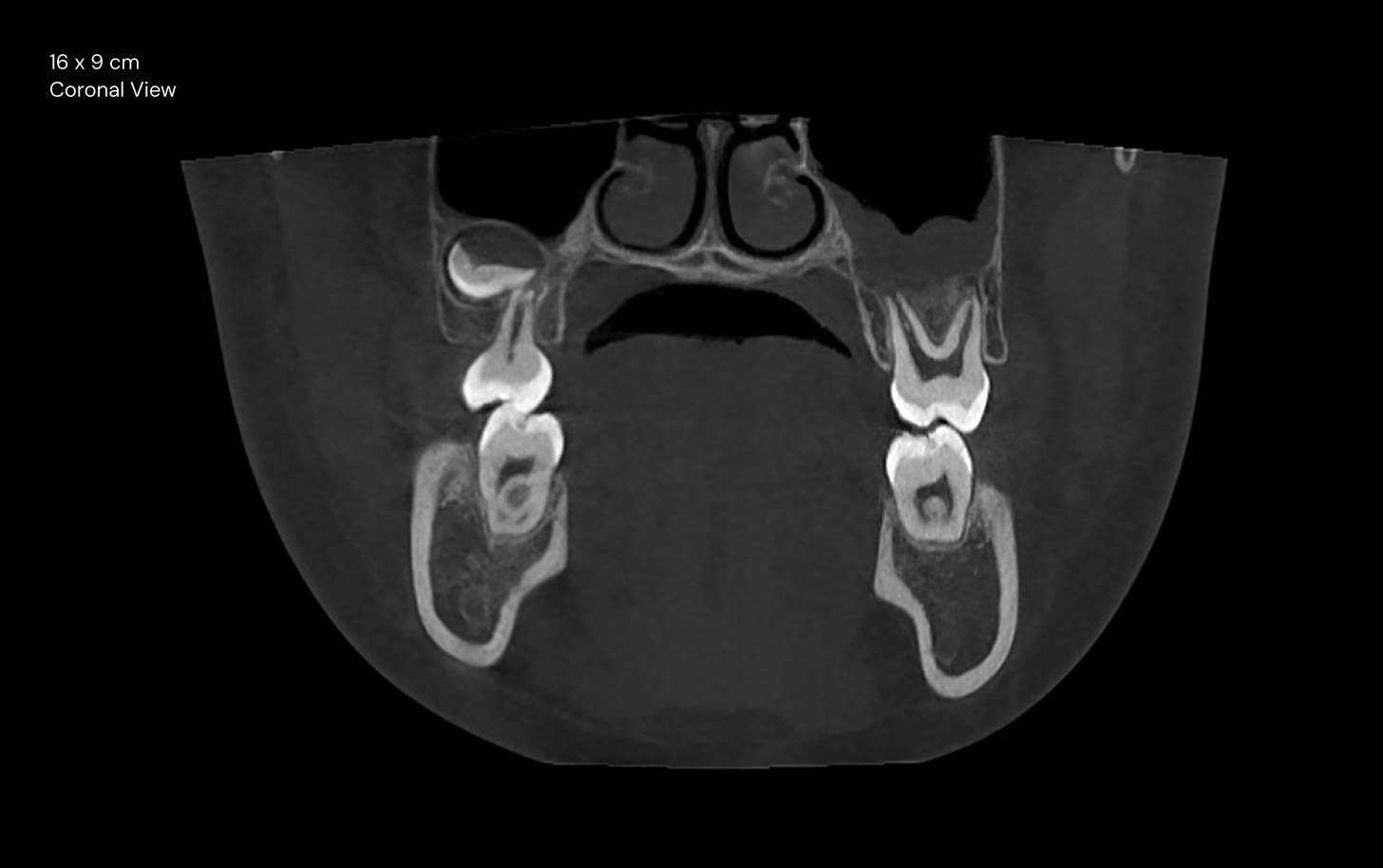
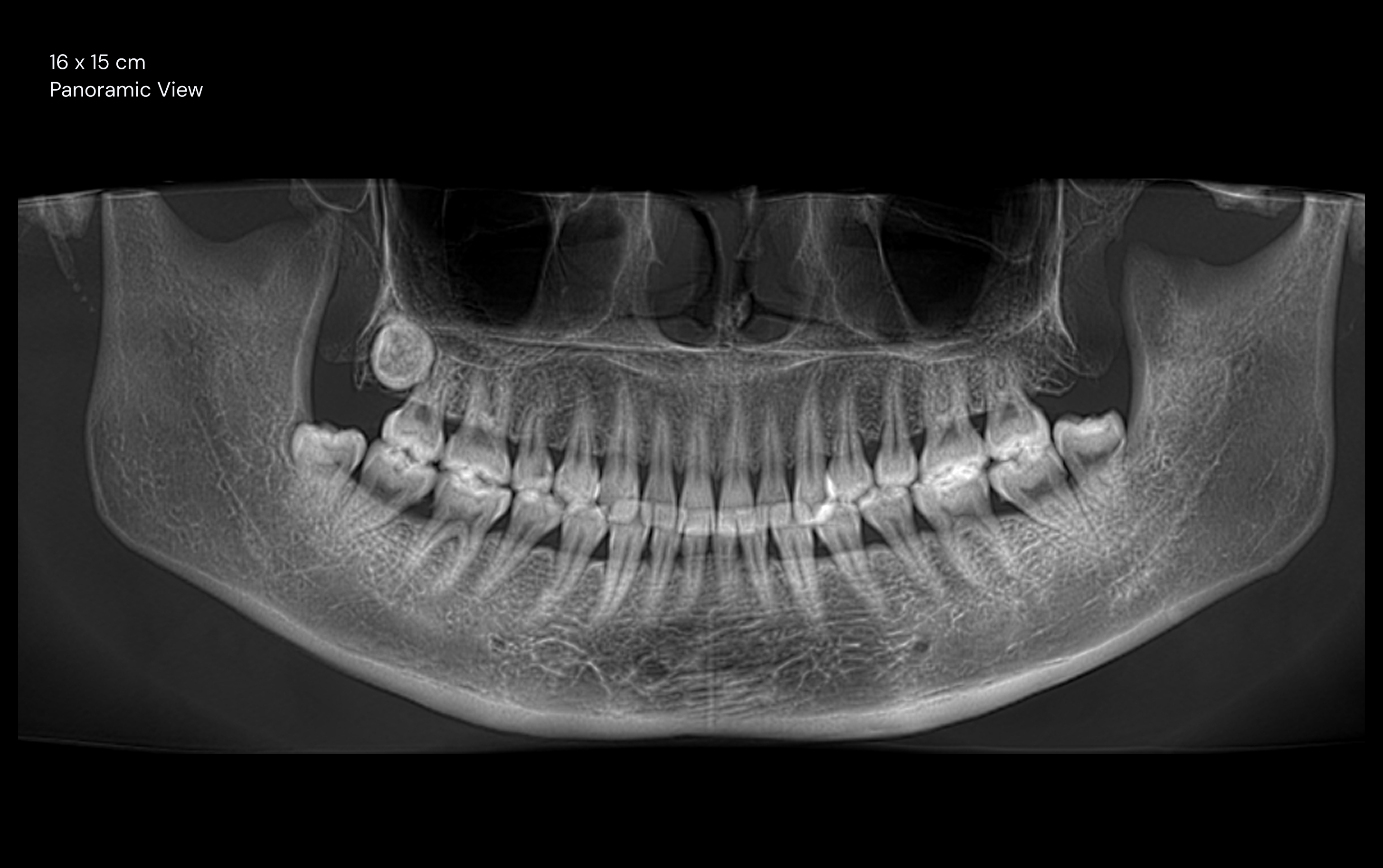
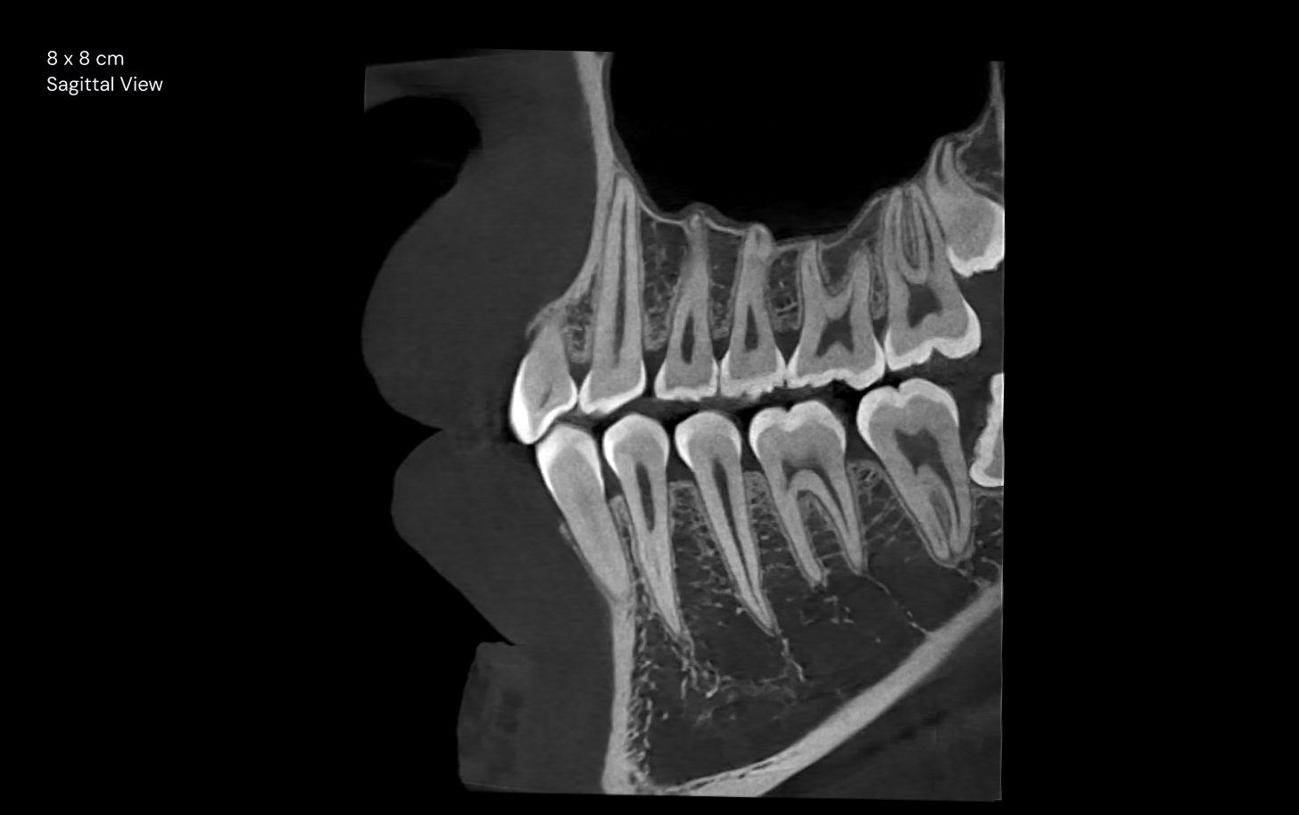
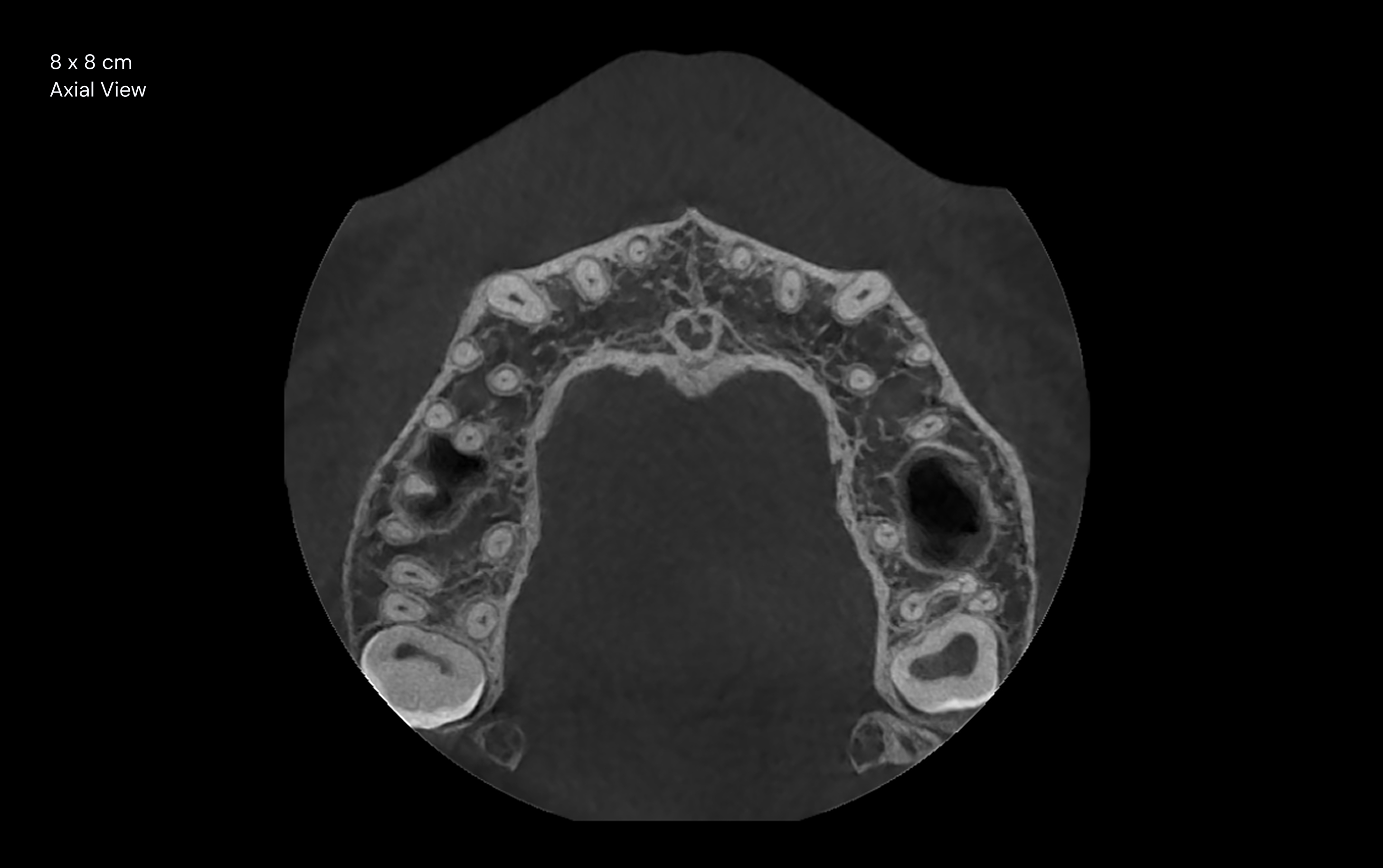
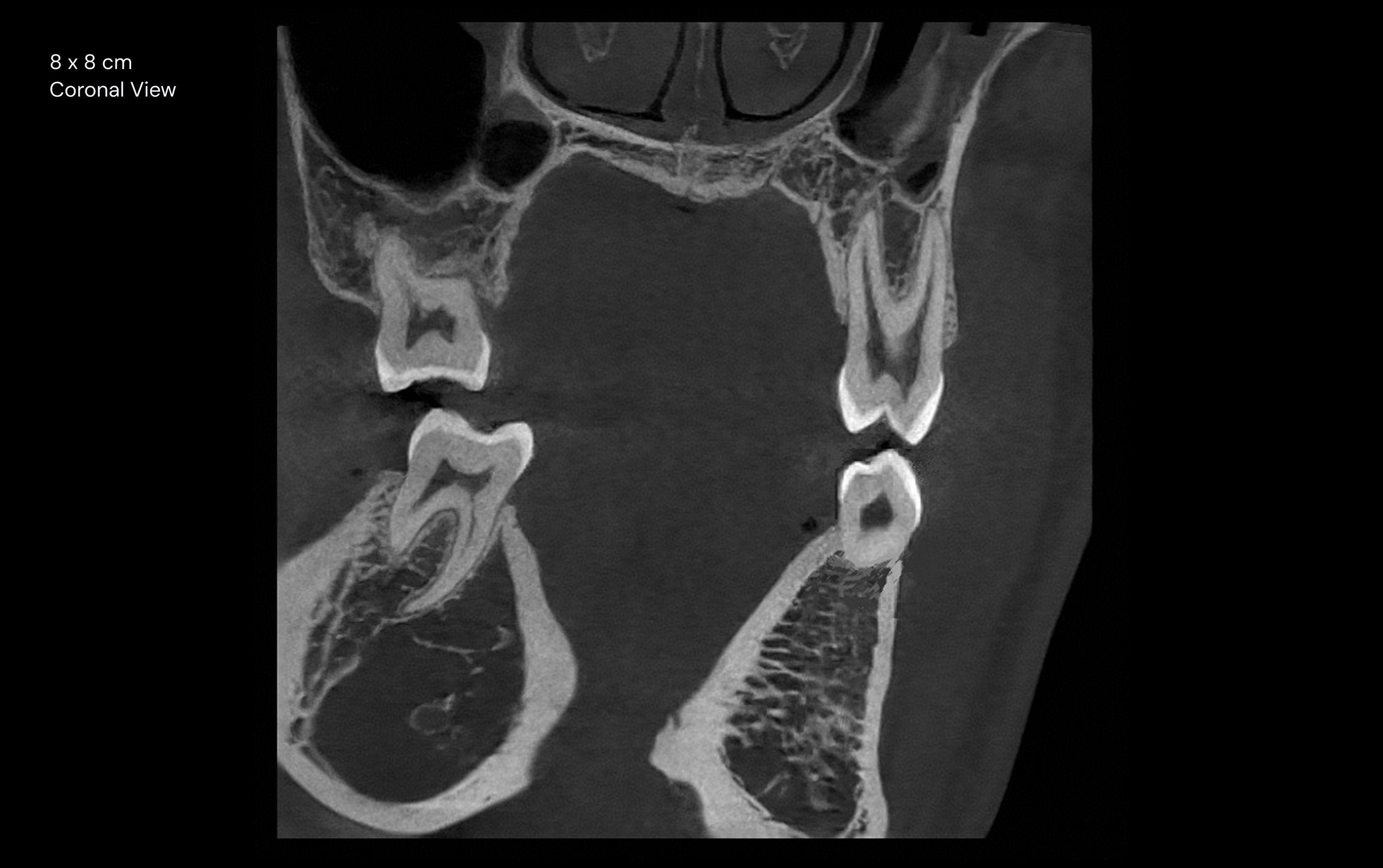
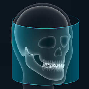
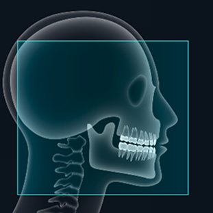
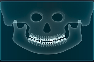
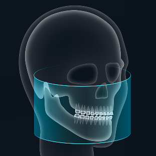
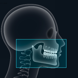
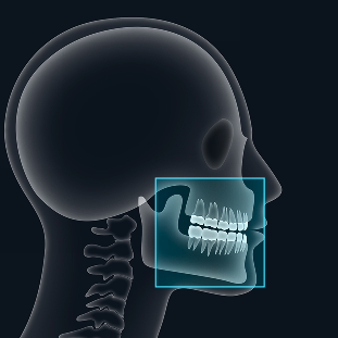
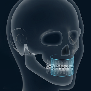
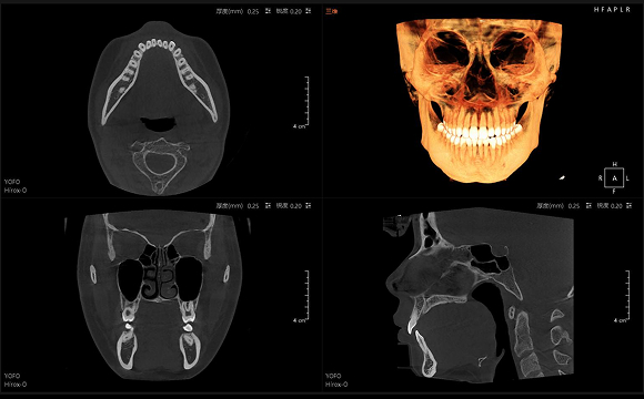
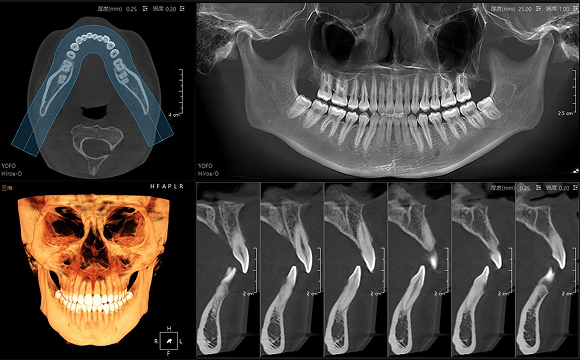
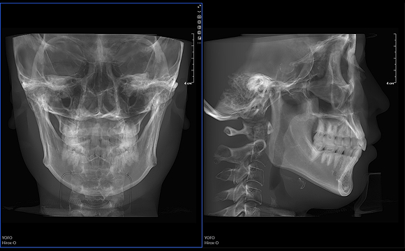
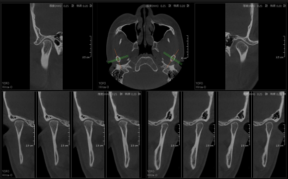
Acquire CT, digital panoramic, 3D frontal and lateral views and 3D TMJ views simultaneously from a single rotation scan.
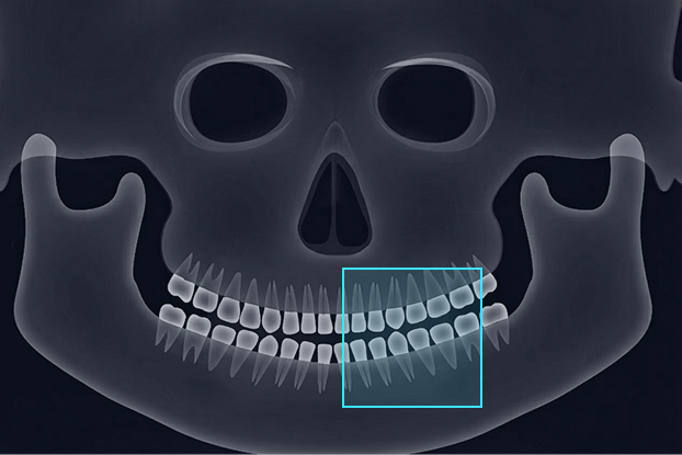
A True 16 × 15 cm Large FOV
FOV options:
16 × 15 cm / 16 × 9 cm /
8 × 8 cm / 4 × 4 cm
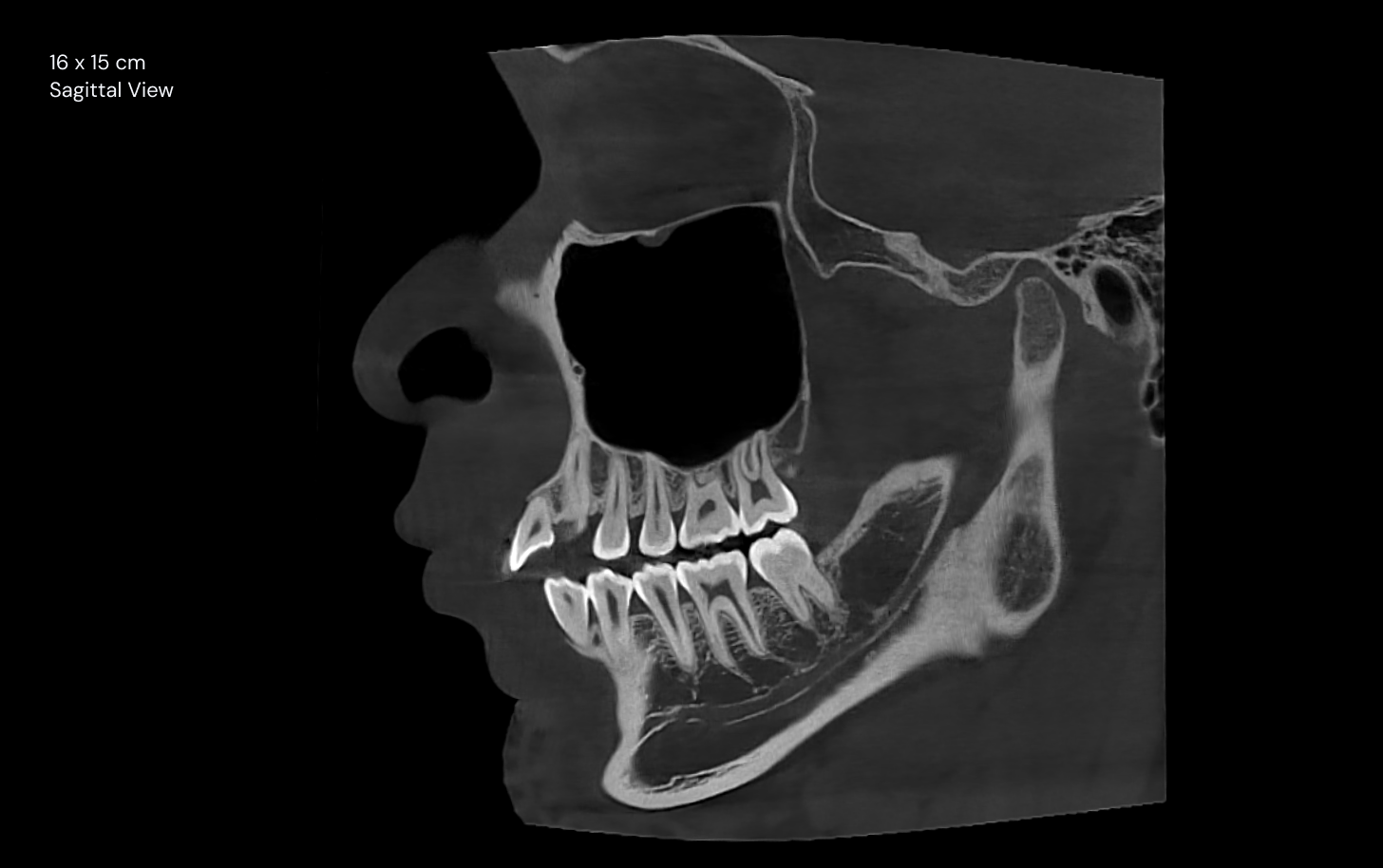
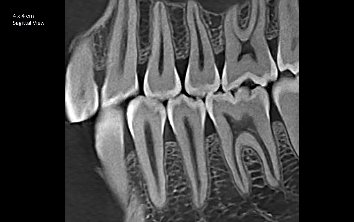
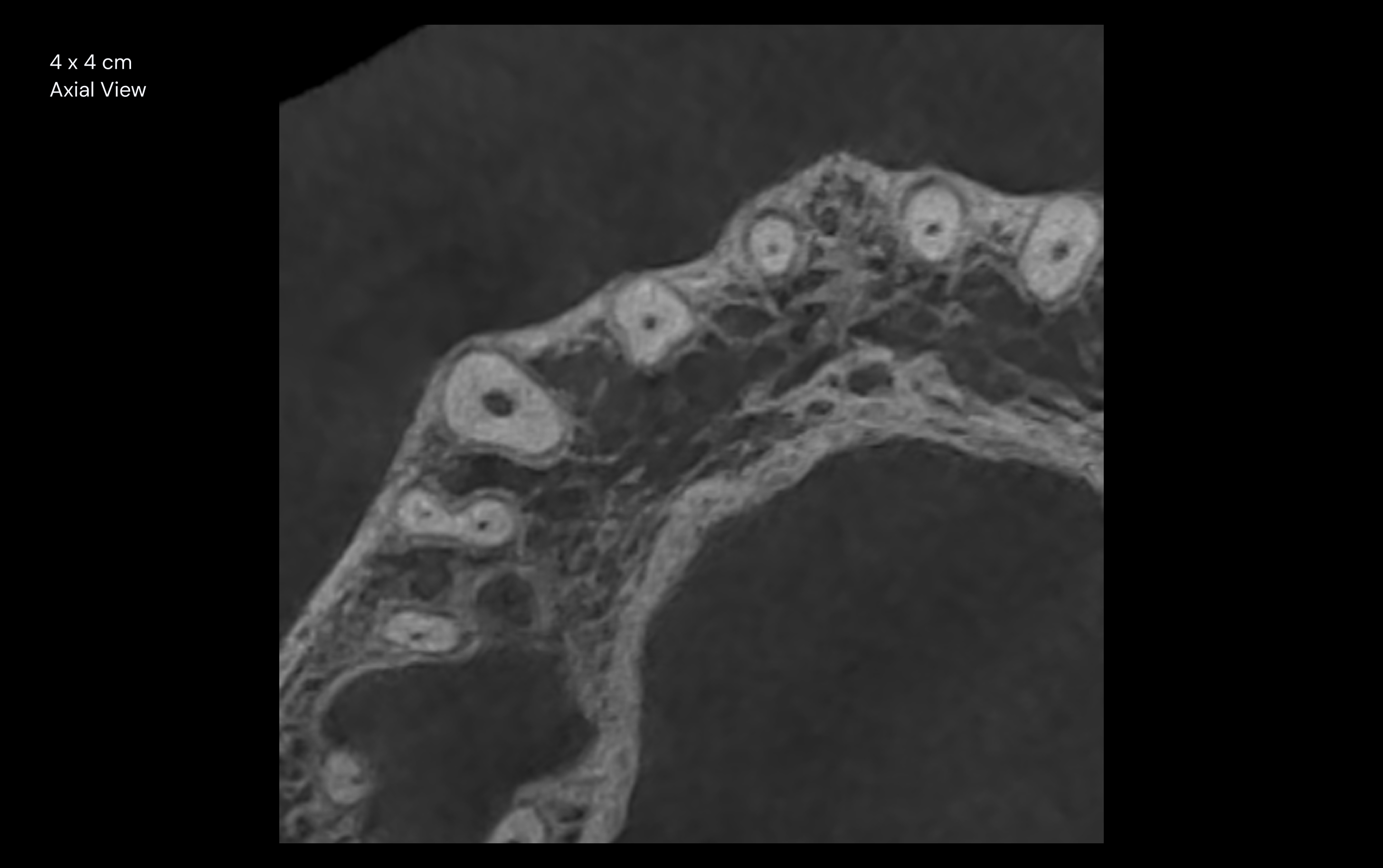
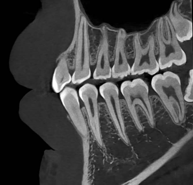
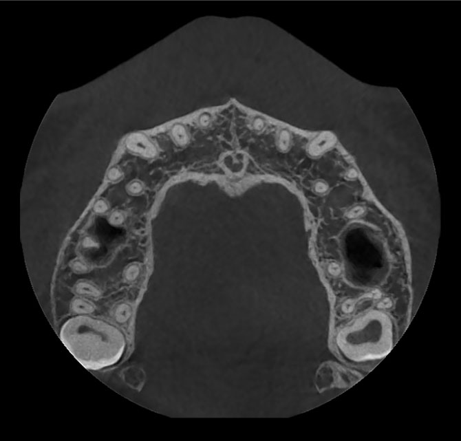
25 lp/cm
Spatial Resolution
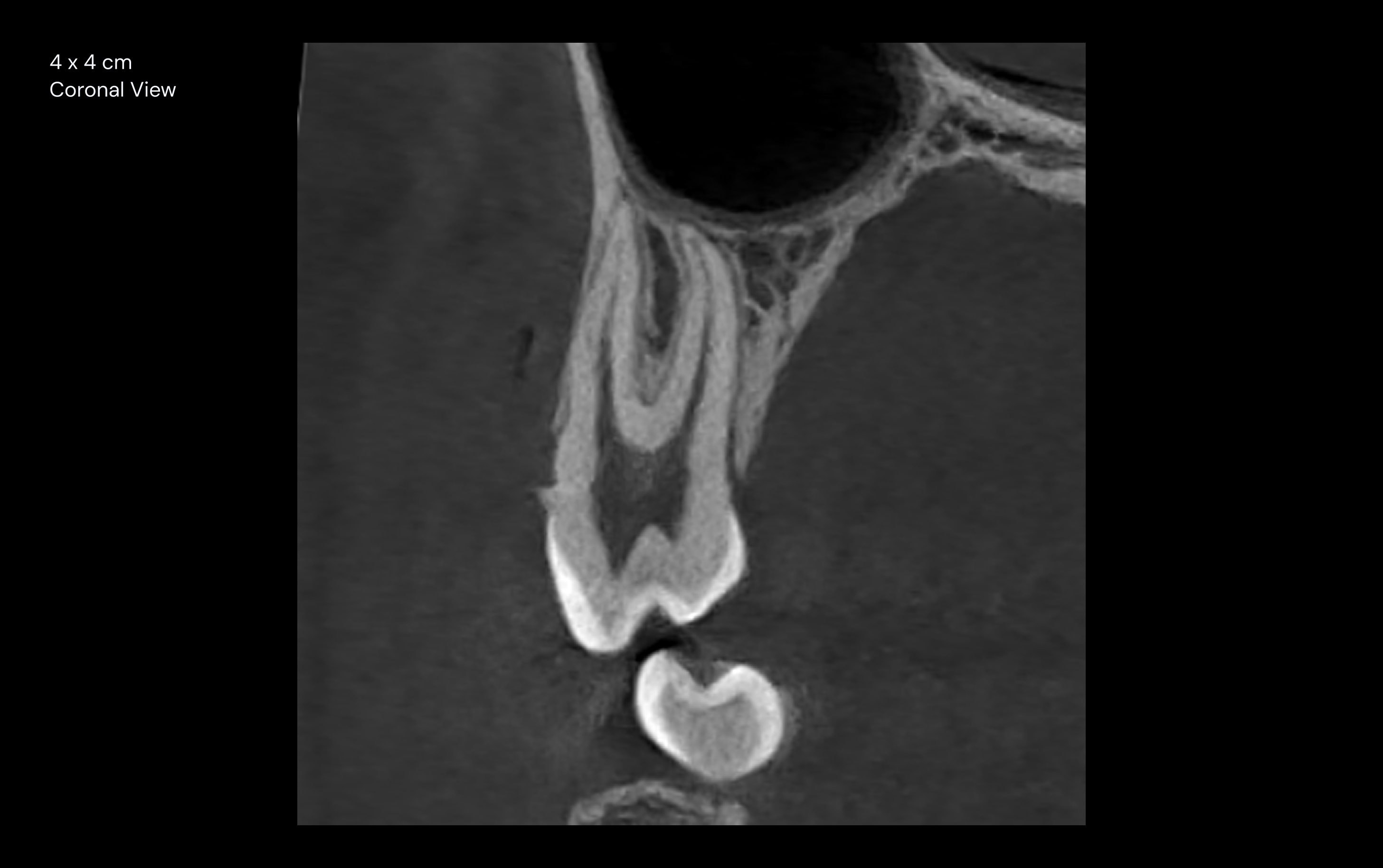
Fine anatomical structures with high clarity.
2.6 s Image Reconstruction
Large-FOV CT images are reconstructed in
just 2.6 seconds.
25 lp/cm Spatial Resolution
Fine anatomical structures are clearly visualized.
One Scan. Everything You Need.
Acquire CT, digital panoramic, 3D frontal and lateral views and 3D TMJ views simultaneously from a single rotation scan. Improve workflow efficiency, reduce unnecessary repeat examinations and minimize patient radiation exposure.
CTPanoramic3D Frontal & Lateral3D TMJ
Designed Around Patients and Practices
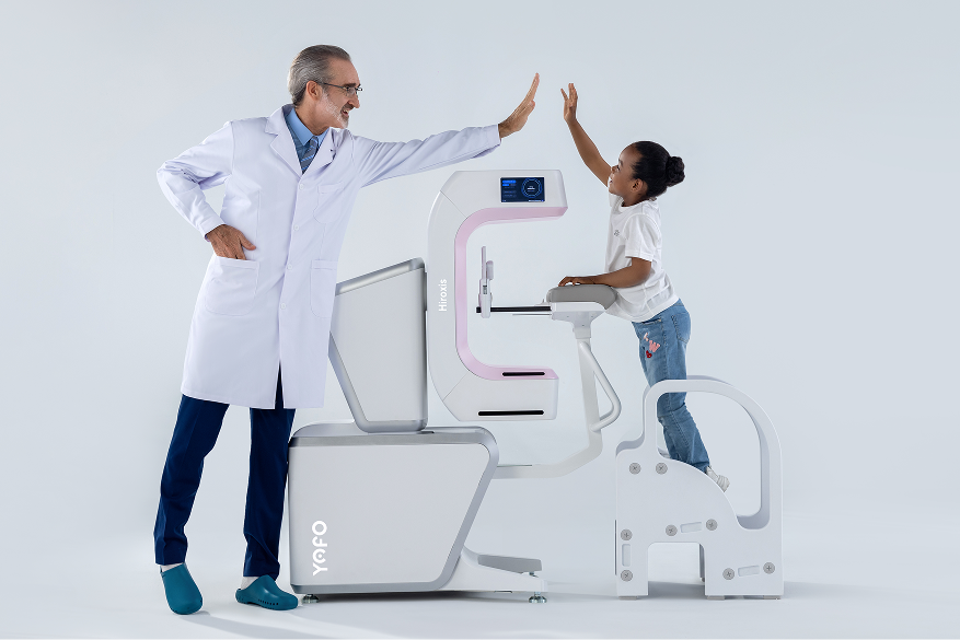
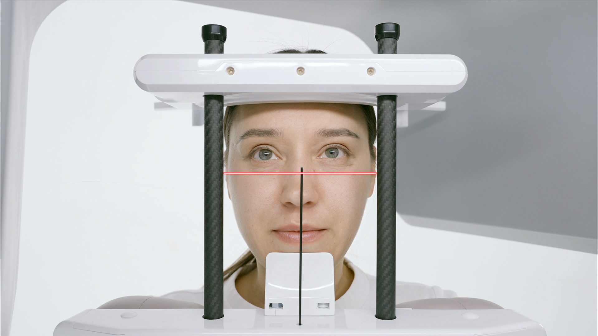
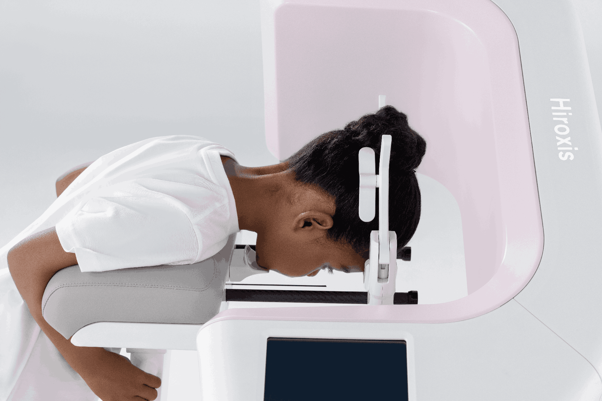
Stable positioning helps children, older patients and other patients remain still during scanning, reducing repeat scans and unnecessary radiation exposure.
Shoulder rests support patient stability, enable quick and easy setup and minimize motion artifacts
Faster positioning streamlines operator workflow and improves overall clinical efficiency.
Consistent patient positioning delivers more reliable image quality across operators.
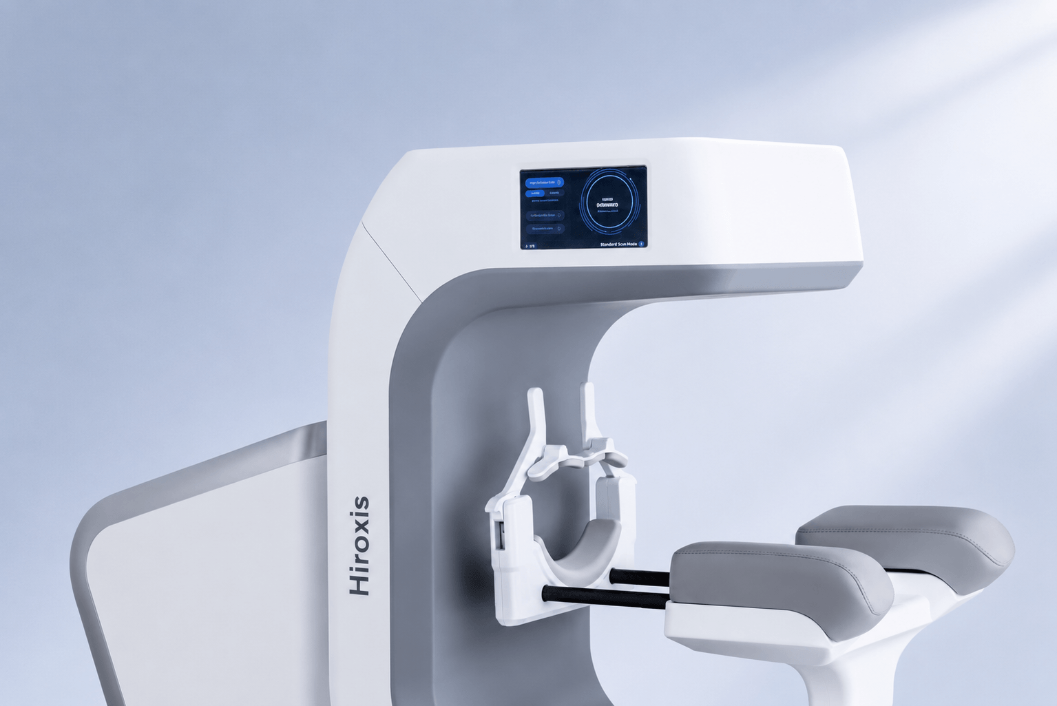
The compact footprint supports flexible room planning, making Hiroxis ideal for practices where space matters.
Built to Last. Designed for Value.
Engineered for up to 12 years of reliable operation, Hiroxis delivers exceptional durability and a lower total cost of ownership
through two key engineering innovations.
Minimal X-ray Tube Wear
Low operating current and short pulsed exposures help maximize X-ray tube life for reliable long-term performance.
4–8 mA typical operating current for high-quality imaging with reduced tube wear.
≤20 s pulsed exposure minimizes operating time and extends tube life.
Reliable Mechanical Design
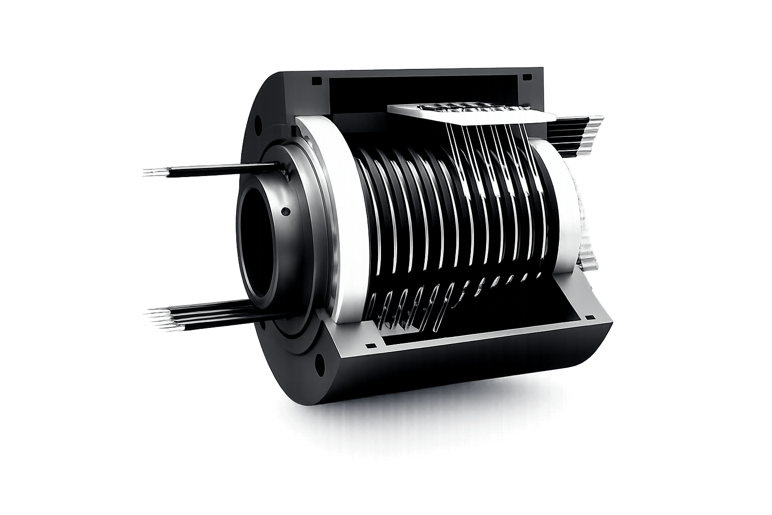
An integrated structure and slip-ring technology help maximize system durability for reliable long-term performance.
A one-piece integrated design reduces movement between components for stable imaging performance.
Slip-ring data transmission minimizes mechanical wear for greater reliability and longer service life.
Refined Industrial Design
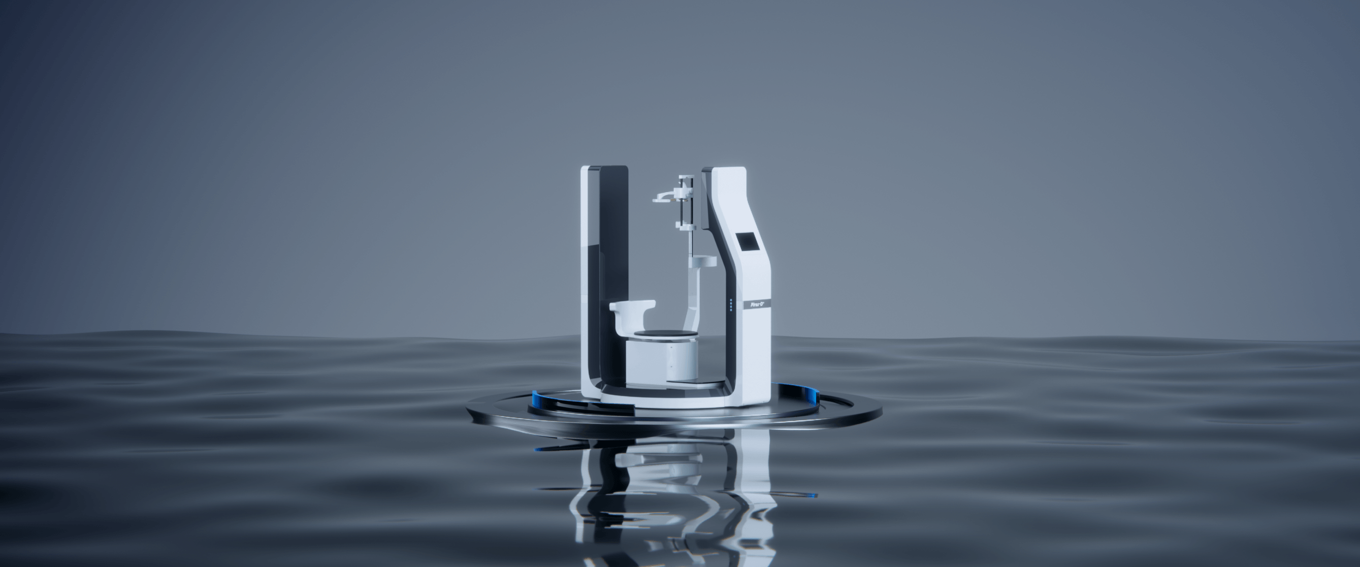
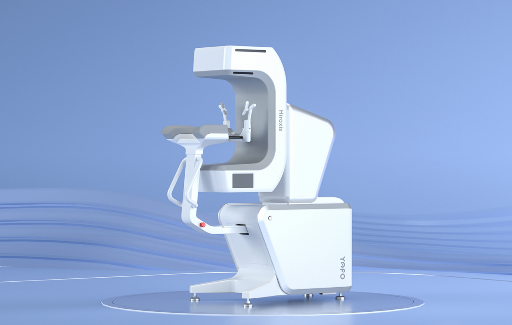
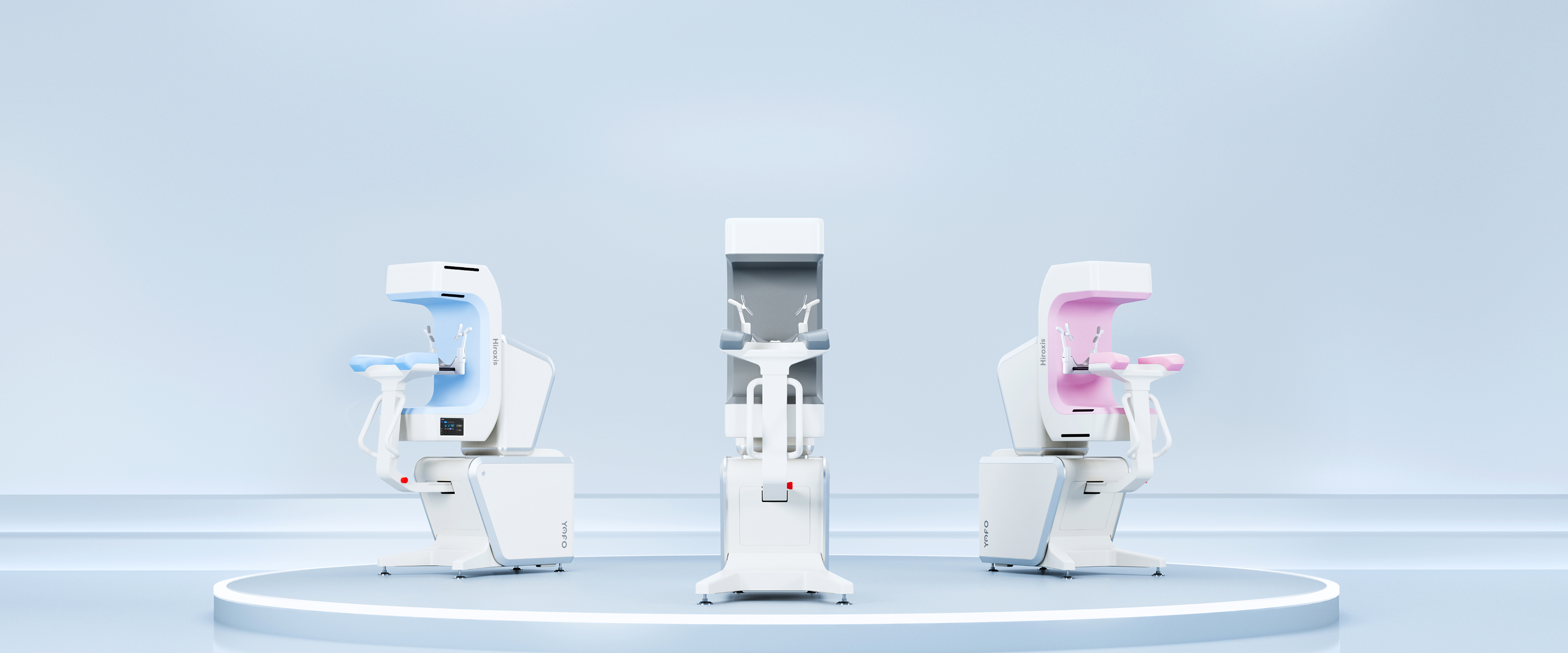
Moving beyond conventional CBCT aesthetics, Hiroxis features a distinctive industrial design that stands out in any clinical environment. Customizable color options provide greater flexibility for different practice settings.
Technical Specifications
Scanning Mode
Field of View (FOV)
16 × 15 cm / 16 × 9 cm / 8 × 8 cm / 4 × 4 cm
Imaging Mode
Simultaneous Multi-View Acquisition
Imaging Parameters
Grayscale
16-bit
Maximum Spatial Resolution
Up to 25 lp/cm
Voxel Size
0.06–0.25 mm
Scan Time
12 s, 20 s, 26 s
Reconstruction Time
2.6 s
X-ray Source
Tube Voltage
60–90 kV
Tube Current
2–12 mA
Focal Spot Size
0.5 mm
Detector
Detector Type
Indium Gallium Zinc Oxide Flat Panel Detector
Dimensions
System Dimensions
594 × 1122 × 1450 mm (±15 mm)
23.4 × 44.2 × 57.1 in (±0.6 in)
Product Name:
Dental Cone Beam Computed Tomography System
Model:YOFO-Hiroxis-15
Intended Use:
The Dental Cone Beam Computed Tomography System is intended to be used by medical institutions for X-ray image diagnosis on oral and maxillofacial areas through X-ray Cone Beam Computed Tomography (CBCT).This product is reusable.
Intended users:
Radiologists or technologists with a background in medical imaging in medical institutions
Contraindications:
The system is not recommended for pregnant women or women who may become pregnant.
*For prohibited contents or precautions, please refer to the instructions. Please read the product instructions carefully or purchase and use under the guidance of medical professionals.
X-ray Radiation safety:
This system is required to generate X-ray radiation during use to achieve its intended use, while the X-ray radiation may be dangerous. Operators are strongly urged to follow the radiation protection procedures and the relevant requirements of user manual, otherwise the useful and scattered beams can cause harm to patients and operators! The operators and maintenance personnel of this system should be properly trained. The Company believes that all authorized operators and maintenance personnel of this system should understand and have adequate training and knowledge on the dangers of X-rays. The Company is not responsible for any injuries or damages caused by X-ray radiation.
Electric Shock safety:
When the system is on, the components carry dangerous voltages, and if there is any improper use and maintenance, it may cause electric shock hazards. To avoid the risk of electric shock, it is strictly forbidden to disassemble the protective enclosure of the components when the equipment is powered on, nor to turn on the device after removing the protective enclosure of the components; It is strictly forbidden to clean the components when the system is powered on. To avoid the risk of electric shock and affecting the basic performance of the system, do not use a damp cloth to clean the inside parts of the equipment, and prevent from liquid entry inside when cleaning the surface of the equipment. To avoid the risk of electric shock, this equipment must only be connected to a supply mains with protective earth.
This website stores cookies on your computer. These cookies are used to collect information about how you interact with our website and allow us to remember you. We use this information in order to improve and customize your browsing experience and for analytics and metrics about our visitors both on this website and other media. To find out more about the cookies we use, see our Privacy Policy.
If you decline, your information won’t be tracked when you visit this website. A single cookie will be used in your browser to remember your preference not to be tracked.
Cookie Settings
Strictly Necessary
Always Active
These cookies are necessary for the website to function securely and cannot be switched off through our Cookie Settings.
They are usually set in response to actions you take, such as setting your privacy preferences, submitting forms or accessing secure areas of the website.
You can set your browser to block or alert you about these cookies, but some parts of the website may not function properly.
Analytics
These cookies help us understand how visitors use our website, including which pages are visited, how visitors navigate through the site and how our website performs.
The information helps us measure and improve website performance, content and user experience.
If you do not allow these cookies, we will have less information about how our website is being used.
Functional
These cookies enable enhanced functionality and help us remember choices you make, such as language, region or other website preferences.
They may be set by Yofo or by third-party services used on our website.
If you do not allow these cookies, some features may not function as intended.
Advertising & Targeting
These cookies may be used to measure the effectiveness of our marketing and provide advertising that is more relevant to your interests.
They may be set by Yofo or by advertising and marketing partners and may recognise your browser or device across other websites.
If you do not allow these cookies, you may still see advertising, but it may be less relevant to your interests.



_1.png)
_1.png)
_2.png)
_3.png)


 Faster positioning streamlines operator workflow and improves overall clinical efficiency.
 Consistent patient positioning delivers more reliable image quality across operators.
 Improved patient stability helps reduce motion artifacts, minimizing repeat scans and unnecessary radiation exposure.
 The compact footprint supports flexible room planning, making Hiroxis ideal for practices where space matters.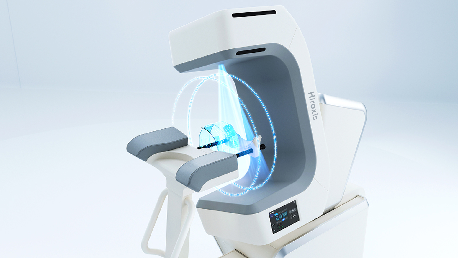

 Scanning Mode
 Imaging Parameters
 X-ray Source
 Detector
 Dimensions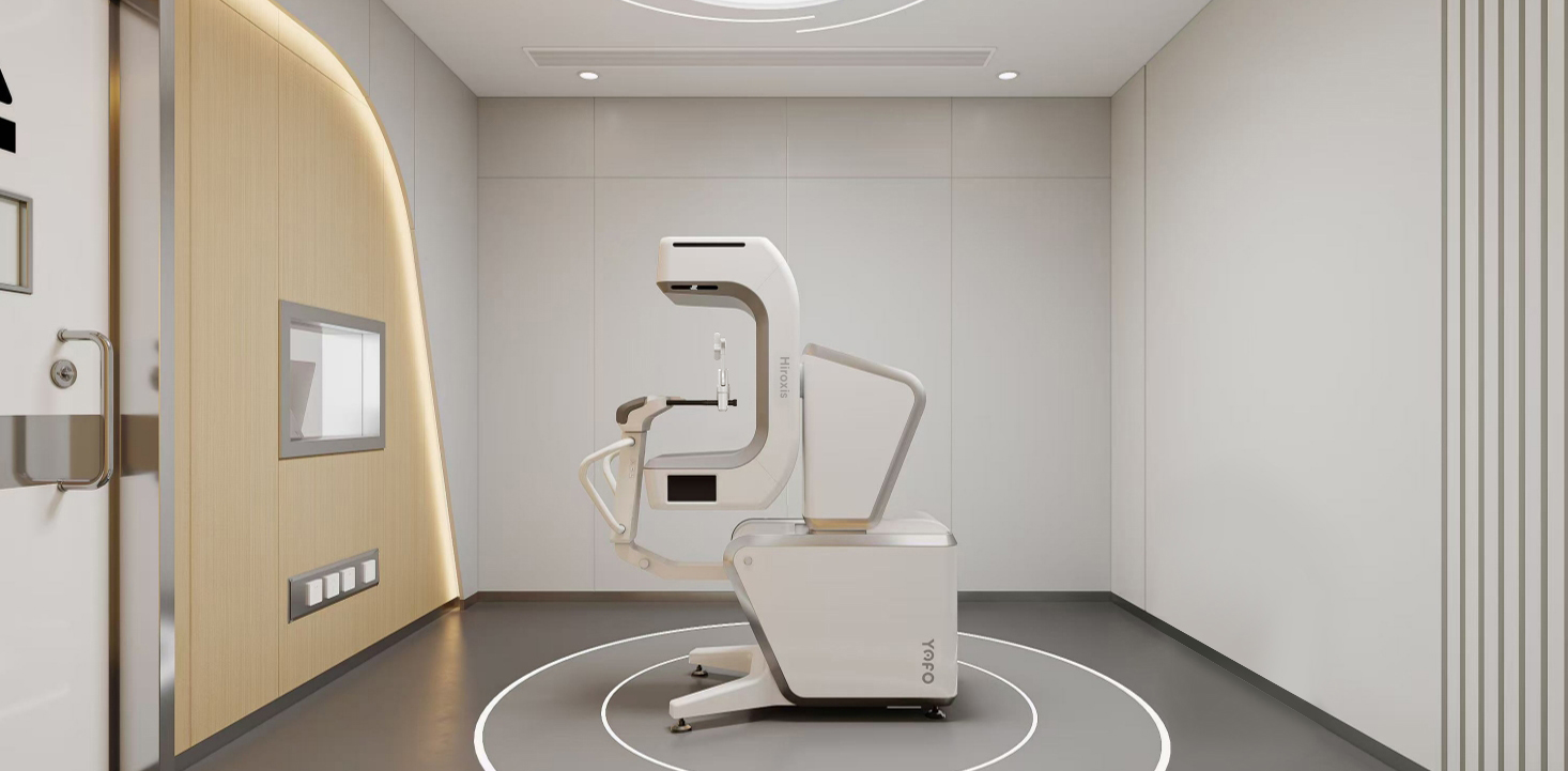
_1.png) 40%
40% 80%
80%_1.png) 300+
300+ 20+
20+ 1,000+
 NO.1
NO.1





_1.png)